Radiologic Science Students Present Research on New Advancements in Medical Imaging
Artificial intelligence, or AI, is leading to a transformation in how work is conducted in many professional fields, including medicine.
There are some who are worried AI may take over the jobs of people. However, students in Holy Family University’s Radiologic Science Program believe AI and other medical advancements are not something to fear, but to embrace.
On November 14, second-year Radiologic Science students presented research posters at the Center for Teaching & Learning in the Library. The students were divided into groups of two or three to explore advancements in seven different imaging modalities: CT, MRI, mammography, radiation therapy, nuclear medicine, sonography and interventional radiology.
Holy Family’s Radiologic Science program is a 20-month program that offers students an Associate’s degree and enables them to take the national certification examination in Radiography through the American Registry of Radiologic Technology. The degree and certification allows graduates to work in several healthcare settings, including hospitals, outpatient centers, physician offices, research and more.
Instructor Jessica Collins, B.S.R.S, R.T. (R) (ARRT), said the poster research project provides students an opportunity to explore potential post-graduation career options.
“It’s a great project for them to review all of the modalities, come up with a plan, and see what each modality has to offer,” Collins said.
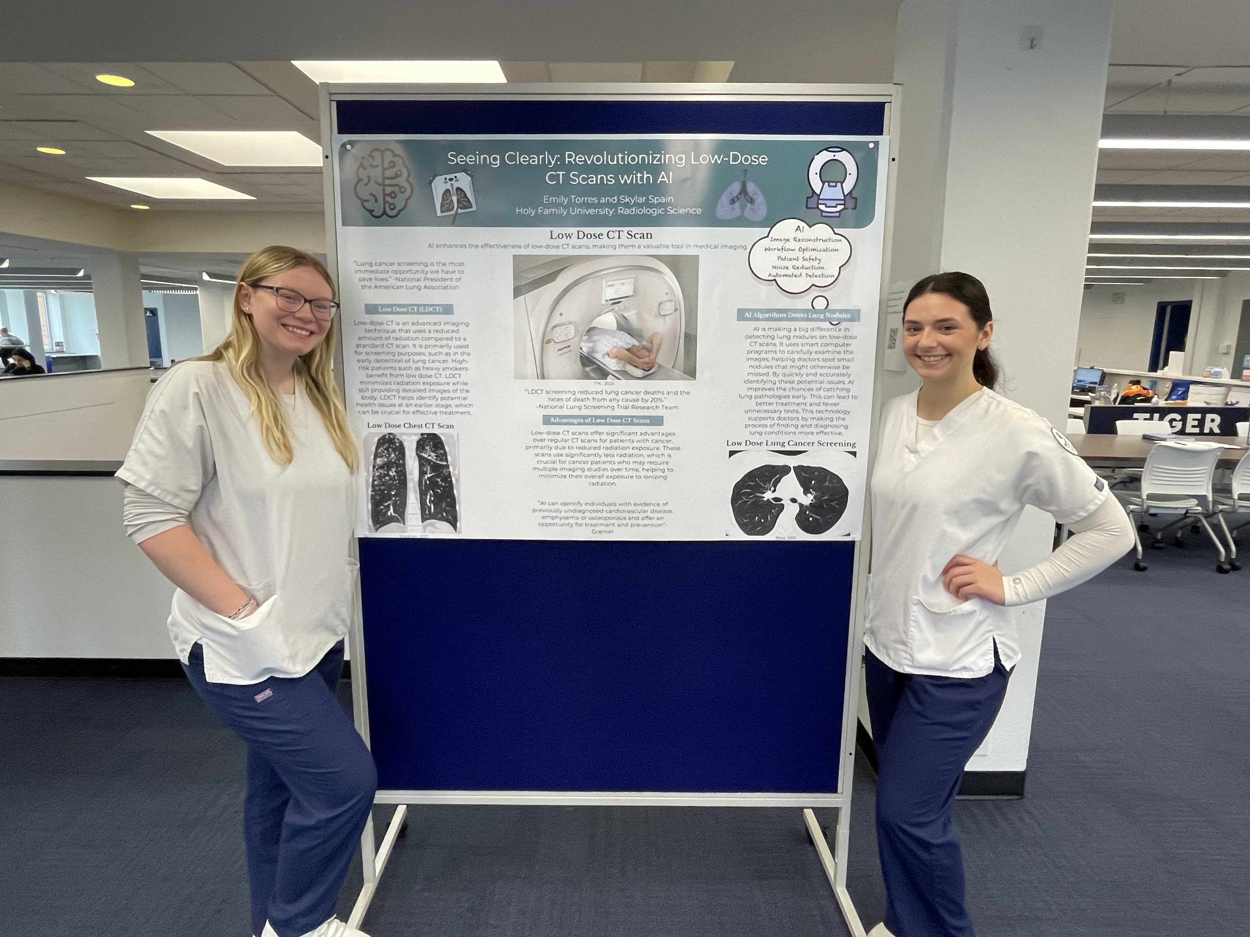
One of the key messages students discussed was how imaging advancements and AI will act as supplementary tools for healthcare professionals in the future. Emily Torres and Skylar Spain discussed how AI can enhance the efficacy of low-dose CT scans as a screening tool for lung cancer.
“AI algorithms help detect nodules quicker and easier with less radiation exposure,” Torres said.
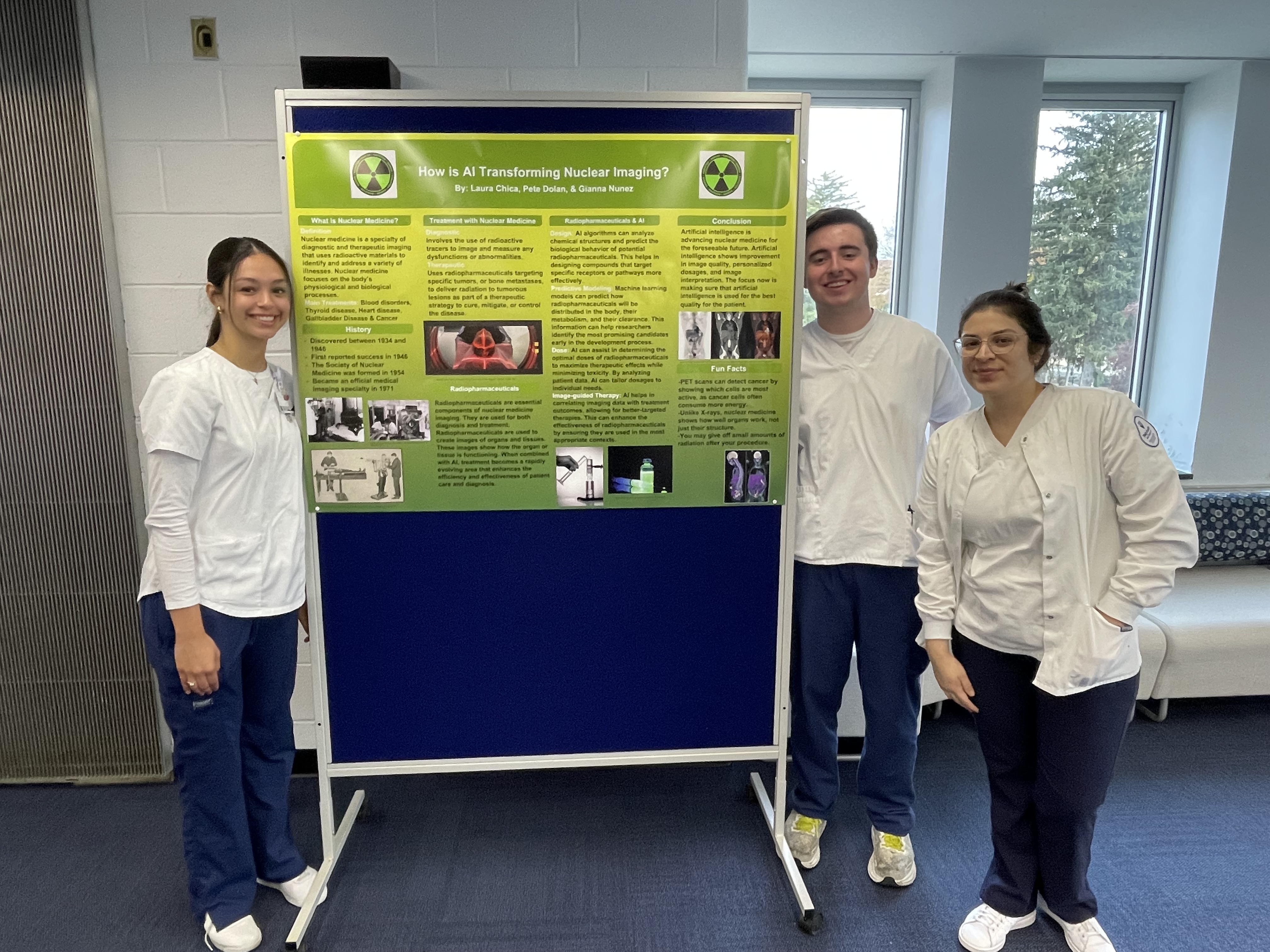
In nuclear imaging, Laura Chica, Pete Dolan and Gianna Nunez discussed ways AI algorithms can better individualize care for radiopharmaceutical therapies and provide treatment recommendations through imaging analysis, which could improve patient outcomes for cancer, heart disease, blood disorders and more.
“AI serves as a helping hand in the whole process,” Dolan said.
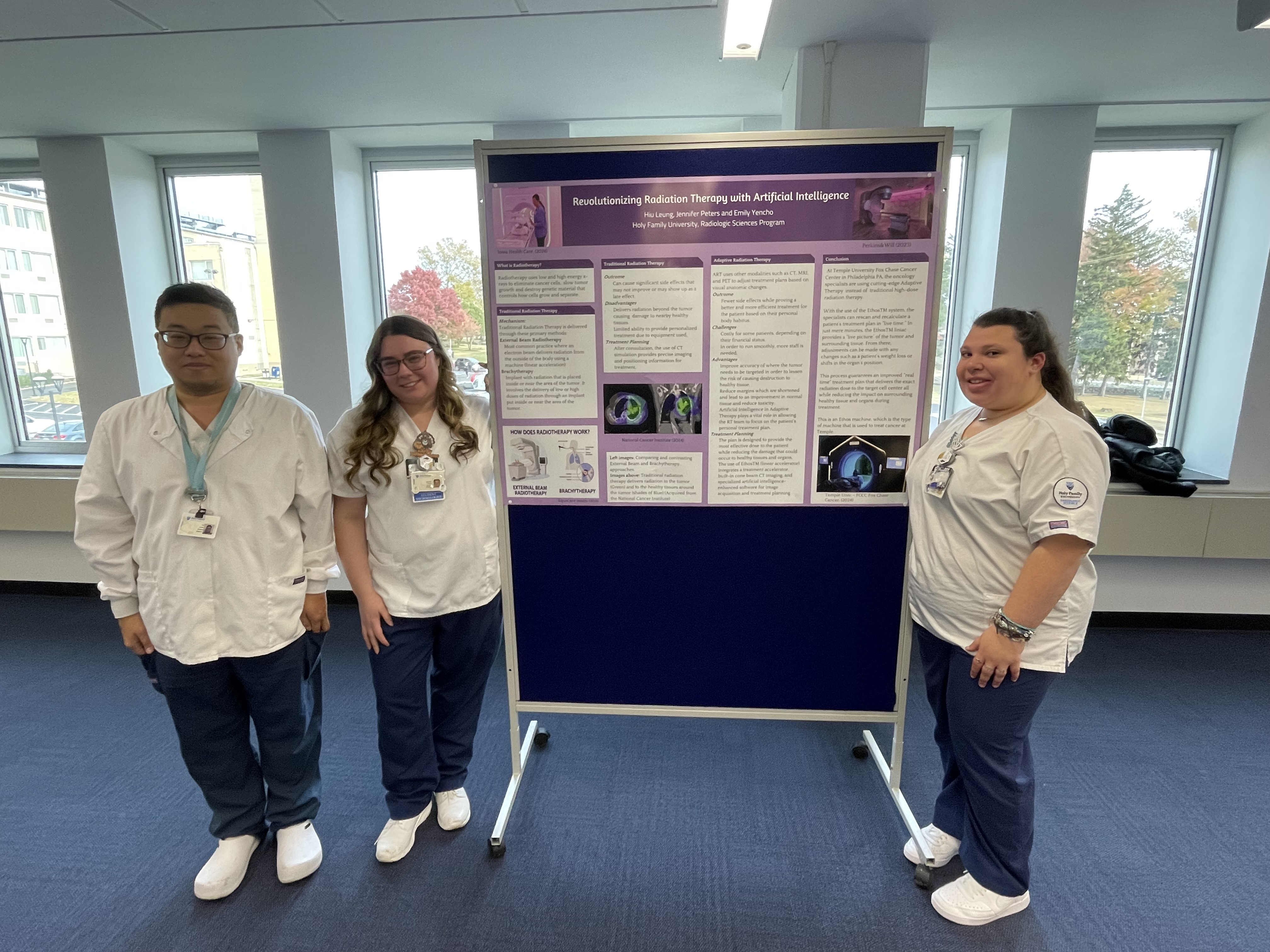
Individualized care is also being used to transform traditional radiation therapy to treat cancer. Hiu Leung, Jennifer Peters and Emily Yencho discussed how adaptive radiation therapy with the help of AI can allow specialists to adjust radiation treatment to focus on the part of the body where it is needed the most. The goal is to reduce side effects and make treatment more efficient for patients.
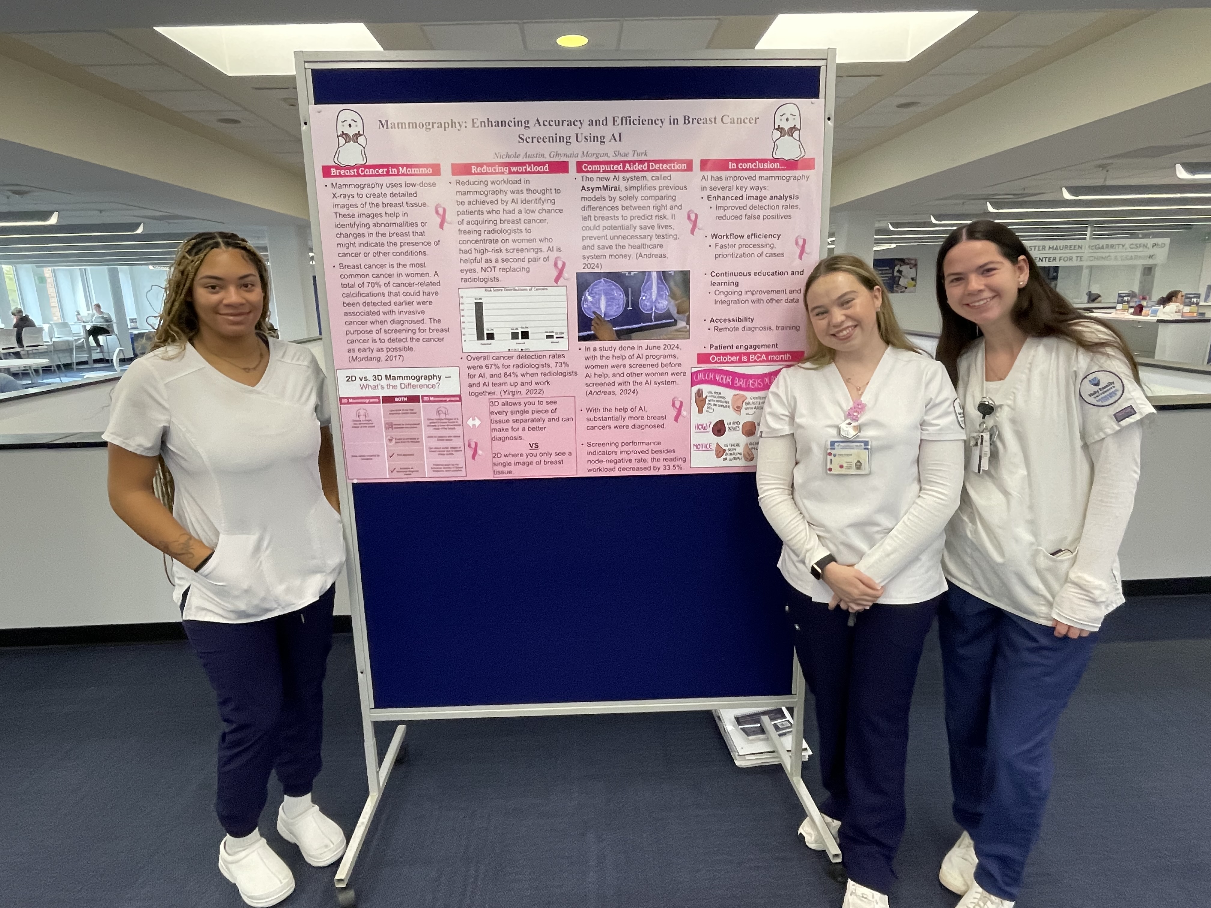
AI is also improving breast cancer detection rates during mammograms. Ghynaia Morgan, Nichole Austin and Shae Turk reported on how AI helped improve breast cancer detection rates. Austin cited a study where cancer detection rates were 84% for radiologists using AI compared with 67% for radiologists not using AI and 73% for AI alone.
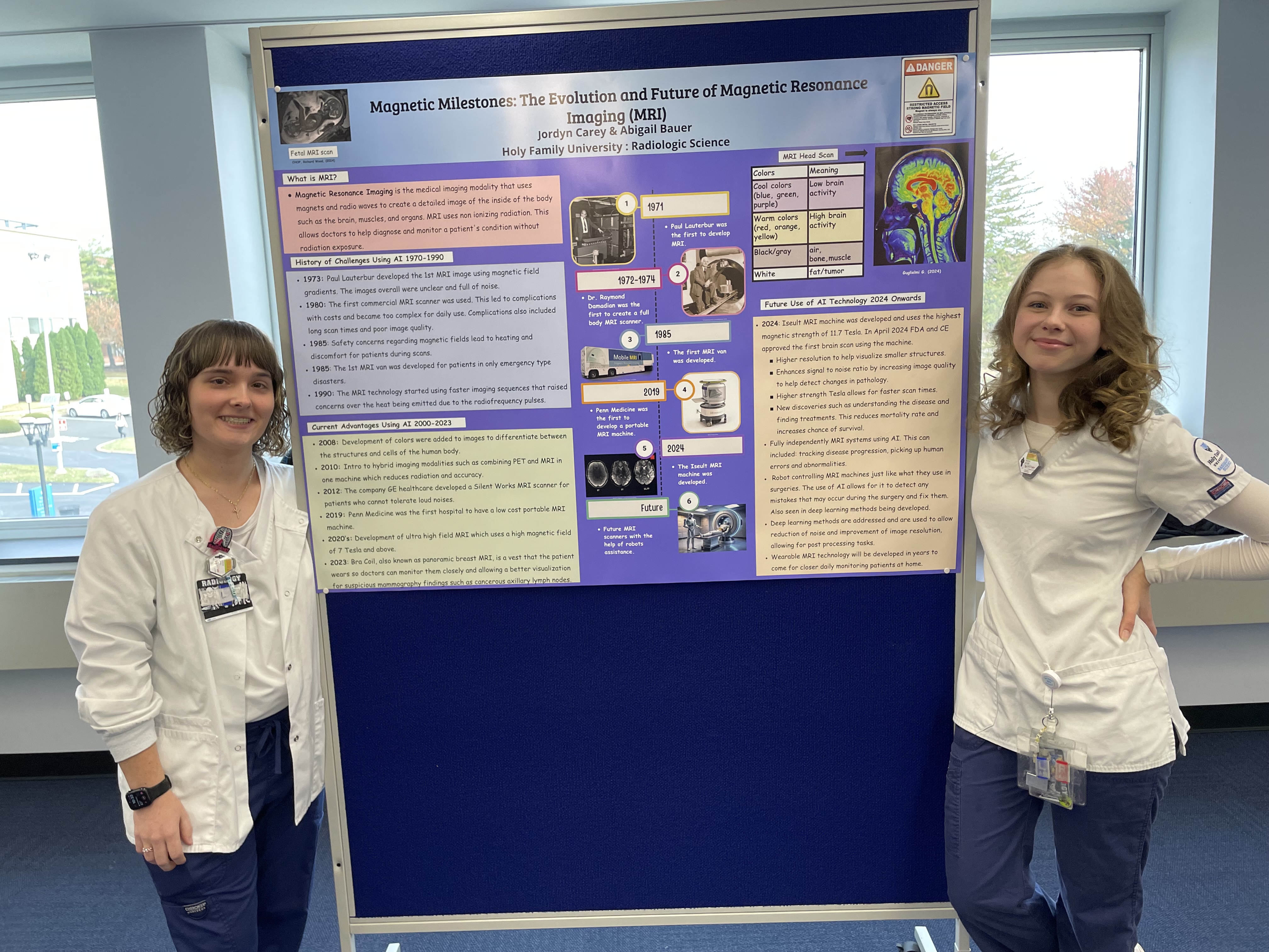
Jordyn Carey and Abigail Bauer’s research explored the evolution of MRI machines and how new advances and AI will improve the devices in the future. Bauer discussed the recently FDA-approved Iseult MRI machine, which takes higher resolution images, has a faster scan time and may be able to help physicians better understand diseases.
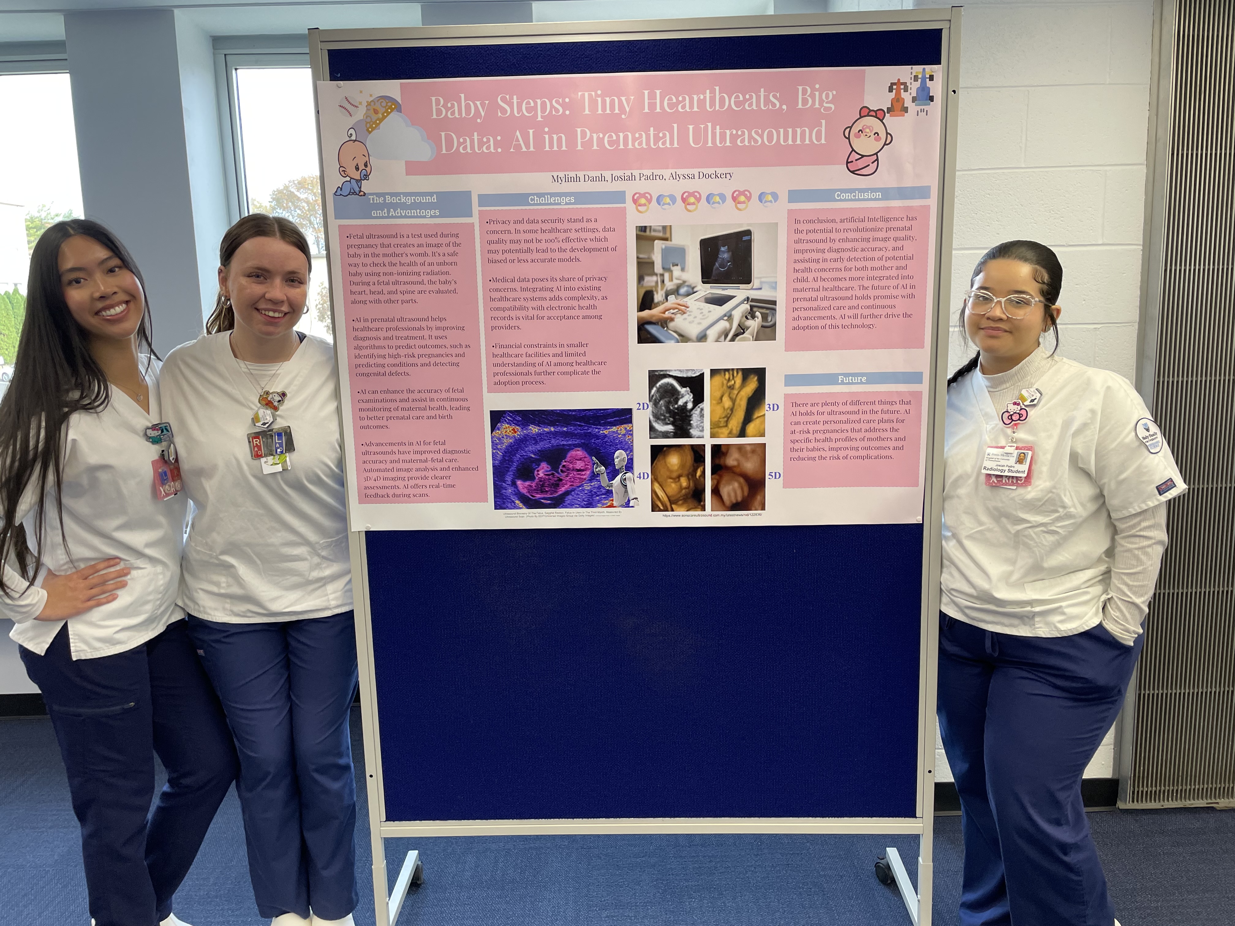
AI is also improving prenatal ultrasounds. Mylinh Danh, Josiah Padro and Alyssa Dockery explored how AI is improving the quality of fetal exams and maternal health by improving ultrasound image quality, diagnostic accuracy and the early detection of possible health issues.
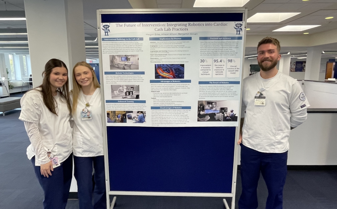
Advances in radiologic sciences could also improve how medical procedures are performed. Megan Biles, Allison Kitson and Benjamin Kirvan explored how robotics is changing the way cardiac catheterizations are performed. Their research showed the use of a robot arm controlled remotely by a surgeon reduces the number of people needed to stand tableside during surgery, lowering the amount of radiation exposure to tableside operators by 95.4%.
